2nd Head of Business Development at ESRF (The European Synchrotron)
Very pleased to be hosting the “CAROTS” (www.carots.eu) network today at ESRF – The European Synchrotron and ILL – Institut Laue Langevin. The project is building a network of intermediaires, like NOVITOM, XPLORAYTION GmbH and SARomics Biostructures AB which help bridge between our amazing Large-Scale European Research Infrastructures and industry, supporting industrial R&D using the exceptional properties of these infrastructures.
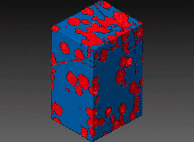
EU_RIs synchrotrons Xrays neutrons
Bernhard Hesse
Jakob Øster
Jacob Becker-Christensen
Selma K.
Solveig Hvidtfeldt
Uwe Sassenberg
Nikolaj Zangenberg
Grethe Jensen
Barbara Fayard
Caroline Boudou
Sophie Bouat
Derek Logan
Thomas Schumann